Jamie Andrews appears to be a rising ‘star’ in the No-Virus movement, gaining prominence over the past month. He claims to have assembled a team with a Contract Research Organization (CRO) to conduct cell culture experiments aimed at invalidating virology. As a result of these experiments, Jamie and No-Virus are declaring the end of virology.
The problem is that Jamie proclaimed the end of virology before releasing any documented evidence of his experiments. To date, he has only released a video with Alec Zeck and Jacob Diaz (a flat-earther) of No-Virus, wherein he falsely claims that his 90+ cell cultures contain different types of viruses without them first being introduced into the cultures to infect cells and replicate.
He has been asking his supporters for donations to conduct nucleic acid sequencing on his samples to determine if they are indeed viruses (hint: they’re not). One would think he would have done this before proclaiming the end of virology.
Other significant problems of this experiment include,
No documentation of methods released.
Has not released the name of the CRO.
Claim cannot be verified because he has not released the methods.
No evidence that any of the particles in his photos are of viruses.
Did no conduct nucleic acid sequencing of his samples to verify his claims.
Here is what I actually wrote:
Here is the image Jamie and No-Virus are calling a vesicle:
The above image is claimed to be a vesicle that is 2000 nm in size. Does this correspond to any normal vesicle in shape, size, or morphology?
Let's review some types of vesicles and their characteristics to see if any align with the structure in the image, which is approximately 2000 nm (2 micrometers) in size.
Types of Vesicles
Exosomes:
Size: Typically 30-150 nm.
Function: Involved in intercellular communication by transporting proteins, lipids, and RNA.
Morphology: Small, uniform, and spherical.
Microvesicles:
Size: Typically 100-1000 nm.
Function: Shed from the plasma membrane and involved in cell signaling.
Morphology: Generally larger than exosomes but still smaller than 2000 nm.
Apoptotic Bodies:
Size: Can range from 500 to 4000 nm.
Function: Formed during programmed cell death (apoptosis) and contain cellular debris.
Morphology: Irregular in shape, containing various cellular components.
Multivesicular Bodies (MVBs):
Size: Can be several hundred nanometers to a few micrometers.
Function: Endosomes containing multiple small vesicles that can fuse with the plasma membrane to release exosomes.
Morphology: Large vesicle containing multiple smaller vesicles.
Vacuoles
Size: Often several micrometers in diameter.
Function: Involved in storage, waste disposal, and maintaining cellular homeostasis.
Morphology: Large, well-defined, and often spherical or irregularly shaped.
Comparison with Image
Given the size (2000 nm or 2 micrometers) and morphology described:
Exosomes and Microvesicles are too small to match the size of the structure in the image.
Apoptotic Bodies can match the size range but tend to be irregular in shape and contain fragmented cellular components.
Multivesicular Bodies (MVBs) could be of a similar size but typically contain multiple small vesicles within a larger vesicle.
Vacuoles are large, well-defined structures that often exceed the size range of typical vesicles and are involved in storage and other functions.
Specialized Large Vesicles
Vesicles larger than 2000 nm are generally not common in typical cellular processes. When they do occur, they are often specialized structures such as:
Large Multivesicular Bodies: These can contain multiple smaller vesicles but are not typically referred to as vesicles themselves.
Giant Unilamellar Vesicles (GUVs): These are synthetic and used in research, not naturally occurring within cells.
Apoptotic Bodies: These can be larger but are specifically associated with cell death and have distinct features such as fragmented cellular components.
Summary
The structure in the image most closely resembles a vacuole due to its size and morphology. While some vesicles, like apoptotic bodies or MVBs, can approach the size range of 2000 nm, they typically have different characteristics (e.g., irregular shape for apoptotic bodies, multiple smaller vesicles within MVBs) that do not match the description or image they claim is a vesicle.
While vesicles can vary in size and function, vesicles larger than 2000 nm (2 micrometers) are quite rare and typically represent specialized forms of extracellular vesicles.
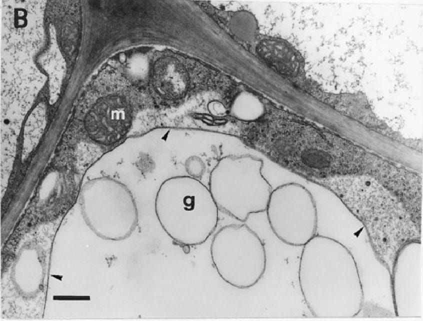
Here is a micrograph of a vacuole (g and surrounding vacuoles):
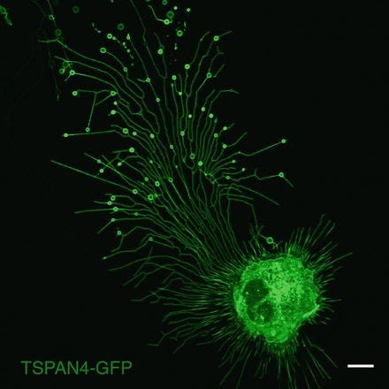
Here is an image of much larger vesicles, migrasomes, as seen at the end of the tendrils they form (from 500-3000 nm):
Does this look like the image provided by Jamie et al.? Absolutely not. Why are Jamie and No-Virus fabricating and misrepresenting their experimental results? And why does Jamie et al. insist on attacking those who merely question or challenge their narrative? Could it be to gain attention for their claims and distract from the fact that their science is deceptive?
Aside from these comparisons, it gives rise to reasonable doubt, as they claim viruses are present in their cultures with the same careless attitude. However, all of the viruses they claim exist in their cultures (HIV, measles, coronavirus), are all enveloped viruses with a lipid bilayer that hides their structure. In such cases, nucleic acid sequencing is essential to determine if a virus is present within the lipid membrane. In such cases, morphology alone can be difficult to determine when a lipid bilayer completely disguises any internal matter. Yet, they are proclaiming that these particles are viruses.
Conclusion
Based on the evidence, I believe that Jamie Andrews et al. are misleading their audience and soliciting support to conduct misrepresented and biased “experiments” to make claims without scientific backing. Their actions appear to be an attempt to create a narrative that viruses do not exist and that virology, along with other branches of science, is fraudulent.
The evidence they present is not only insufficient but also distorted and misrepresented. Instead of providing clear, scientifically valid data, they are manipulating their findings to fit a preconceived narrative. For instance, they claim to have found various viruses in their cell cultures without any proper introduction of these viruses to the cultures. This method defies the fundamental principles of virology, where controlled infection and replication are essential to verify the presence of viruses.
Moreover, Jamie Andrews was interviewed by Alec Zeck and Jacob Diaz—the latter being a confirmed flat-earther. This collaboration further undermines their credibility and exposes a severe bias and predetermined outcome, no matter the evidence. In the video, Jamie asserts that their 90+ cell cultures contain different types of viruses, but they make such claims without having performed the necessary nucleic acid sequencing to confirm the identity of these supposed viruses.
As a result, they are now asking their followers for donations to conduct this sequencing, which should have been done before making any claims about their findings.
By prematurely proclaiming the end of virology and declaring other branches of science fraudulent, they are spreading misinformation and fostering distrust in legitimate scientific research. This behavior not only discredits them as people, but also harms public understanding of virology and science further within their audience.
In summary, Jamie Andrews et al. are engaged in a campaign of misinformation for personal gain, utilizing attacks to deflect from their use of distorted evidence and biased experiments that make unfounded claims.
They are misleading their audience by misrepresenting standard practices of virological protocols and procedures and presenting them as factual representations.
Jeff Green


Thank you, Jeff, for focusing on Jamie's misleading content. I noticed he has a Substack (controlstudies.substack.com). It has only one post, presumably on scientific Controls. I think he was trying for a world record on the most misspelled words in a single post. It's all very sloppy. Along the way he dismisses atomic theory and genetics as pseudoscience. The post concludes with a hilarious misunderstanding of a short report describing how a rapid antigen test won't give reliable results if you don't follow instructions.
I've been learning about virology laboratory procedures by reading some of the documents Jamie Andrews references. It doesn't appear he reads much or he would have a better understanding of the subject. For example, I've seen him post an excerpt from this document a number of times: "Cytopathic Effects of Viruses Protocols" from the American Society for Microbiology. He highlights this paragraph:
"The rate of CPE appearance is also a characteristic that can be used to help identify viruses. In general the rule of thumb is that a virus is considered slow if CPE appears after 4 to 5 days in cultures inoculated at low MOI, and rapid if CPE appears after 1 to 2 days in cultures inoculated at low MOI. It is important to note that at a high MOI all CPE can occur rapidly. So all decisions about rate of CPE appearance should be based on the lowest MOI that produces CPE."
Jamie says "Here is the American Society of [sic] Microbiology stating that if ANY appearance of CPE is in the culture before day 5, you have a "virus" in the culture."
But it doesn't state that. Not even close!
It does state, elsewhere in the same document:
"Recognizing CPE and using it as a diagnostic tool requires much experience in examining both stained and unstained cultures of many cell types."
"The best knowledge of viral CPE comes from experience. And control uninfected cells should always be observed to distinguish normal cell changes that occur as cells age from cytopathic effect."
I guess Jamie didn't read that part. It makes me wonder how an automated machine (Countess) can take the place of an experienced microbiologist when examining cell cultures for CPE.